How to Read a Microhematocrit Tube Reader
Hematocrit - a review of unlike analytical methods
INTRODUCTION
This article describes the advantages and disadvantages of the dissimilar methods of measuring hematocrit by discussing the following:
- The components of whole claret
- Indications for measuring hematocrit
- Measuring technologies
i. Microhematocrit
2. Consummate blood cell count
3. Conductivity on claret gas analyzers
4. Calculation of hematocrit - Comparison of the different technologies
This commodity will be followed by some other article describing the different methods of determining hemoglobin.
THE COMPONENTS OF WHOLE Blood
Whole blood is comprised of erythrocytes (the red blood cells or RBCs involved in oxygen transport), platelets and leukocytes (the white blood cells or WBCs involved in the body's allowed defence force). The cells are suspended in the aqueous medium of plasma.
In blood from healthy individuals, erythrocytes constitute the vast majority of cells; the erythrocytes contain hemoglobin (Hb), which gives claret its red color and which has oxygen-binding abilities. Plasma mainly consists of water (approx. 93 %) only likewise of salts, various proteins and lipids equally well equally other constituents, e.g. glucose.
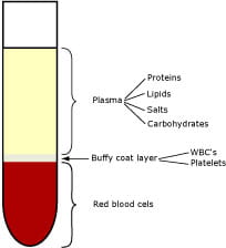
FIG. one. A centrifugated whole-blood sample
The definition of hematocrit (hemato from the Greek haima = blood; crit from the Greek krinein = to dissever) is the ratio of the book of packed red blood cells to the full blood volume and is therefore also known as the packed cell volume, or PCV.
The hematocrit is reported as a percentage or a ratio. In healthy developed individuals the red blood cells constitute approx. xl-48 %, whereas newborns may take hematocrits of up to 60 % [1].
The layer between the RBCs and plasma, the buffy coat layer, constitutes approx. one %. It consists of WBCs and platelets and should therefore not be calculated equally function of the packed jail cell volume.
The human relationship between hematocrit and hemoglobin
The following is a summary of the quantities/abbreviations that are relevant when discussing hematocrit:
- Hct: Hematocrit (% or volume fraction)
- ctHb: Concentration of total hemoglobin (g/dL, g/L or mmol/50)
- RBC: Ruby blood cell (erythrocyte) (× 1012/L)
- MCV: Hateful cell volume (fL)
- MCHC: Mean corpuscular hemoglobin concentration (%, g/L or mmol/L)
In normal conditions there is a linear human relationship between hematocrit and the concentration of hemoglobin (ctHb). An empirical report [ii] has shown that the relationship tin can exist expressed as follows:
Hct (%) = (0.0485 × ctHb (mmol/L) + 0.0083) × 100
Hematocrit tin also be estimated from measurements of the mean prison cell book (MCV) or the mean corpuscular hemoglobin concentration (MCHC):
Hct (%) = MCV × RBC × 0.1
Hct (%) = ctHb × 100
MCHC
INDICATIONS FOR MEASURING HEMATOCRIT
Hematocrit measurements may be requested when it is suspected that a patient is anemic or suffering from dehydration, bleeding or other medical and surgical conditions.
Depression hematocrit
A low hematocrit reflects a depression number of circulating red blood cells and is an indicator of a subtract in the oxygen-carrying capacity or of overhydration. Examples of conditions causing a low hematocrit (anemia) include [iii]:
- Internal or external hemorrhage – haemorrhage
- Complexity of chronic renal failure – kidney disease
- Pernicious anemia – vitamin-B12 deficiency
- Hemolysis – associated with transfusion reactions
A low hematocrit may exist establish in autoimmune diseases and os-marrow failures.
High hematocrit
A high hematocrit may reflect an absolute increase in the number of erythrocytes, or a decrease in plasma volume, in conditions such as [3]:
- Severe aridity – e.g. in case of burns, diarrhea or excessive use of diuretics
- Erythrocytosis – excessive red blood cell product
- Polycythemia vera – abnormal increase of blood cells
- Hemachromatosis – an inherited iron metabolism disorder
Loftier hematocrit is also used every bit an indicator of the excessive intake of exogenous erythropoitin (EPO), which stimulates the production of red blood cells. Athletes tin artificially improve their functioning by enhancing the oxygen-carrying capacity with EPO.
In newborns and peculiarly premature babies, loftier hematocrit values are common. The hematocrit of infants reaches the level of adult hematocrit by approx. three months of age [1].
The atmospheric condition leading to low hematocrit values, east.g. hemorrhage, oftentimes require continuous measurements of the hematocrit and fast decisions apropos transfusions. If the hematocrit is measured immediately afterwards an acute hemorrhage, the value will be normal until the decreased claret volume is corrected past fluid shifts into the blood vessels.
MEASURING TECHNOLOGIES
This section gives a technical description of the about usually used techniques for measuring hematocrit:
- The determination of hematocrit by means of centrifugation
- The calculation of hematocrit from the consummate blood prison cell count (CBC)
- The determination of hematocrit by conductivity
- The calculation of hematocrit from ctHb
1. Microhematocrit
The reference method recommended past NCCLS of determining hematocrit or packed cell book (PCV) is centrifugation.
Method [four]:
Hematocrit (PCV) is the measure of the ratio of the volume occupied by the red claret cells to the volume of whole claret. The claret sample is drawn into a capillary and centrifugated, and then the ratio tin can exist measured and expressed every bit a decimal or percentage fraction.
Materials:
- Whole blood from a freely flowing skin puncture or anticoagulated (EDTA or heparin) venous or arterial claret
- Glass capillary tubes with a narrow diameter
- Sealing compound (if the capillaries are non self-sealing)
- A microhematocrit centrifuge with a maximum relative centrifugal force of 10-15,000 × g, which should be reached within thirty seconds [4]
- Graphic reading device
Procedure:
- Capillary tubes are filled by capillary forces. A minimum of two capillaries is required to ensure remainder in the centrifuge. It is important that the tubes are sealed thoroughly.
- After v minutes of centrifugation the hematocrit can exist measured while the tubes are still kept in a horizontal position. A distinct column of packed erythrocytes is visible in 1 cease of the capillary tube (Fig. 2). The packed erythrocytes are followed by kickoff a pocket-size turbid layer – the buffy coat layer – and then a clear column of plasma (Fig. two). Hematocrit is estimated by computing the ratio of the column of packed erythrocytes to the full length of the sample in the capillary tube, measured with a graphic reading device.
- The measurement should be performed within 10 minutes to avoid merging of the layers.
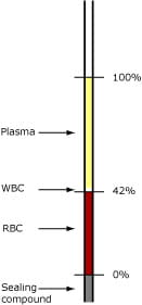
FIG. 2. Reading the hematocrit
Limitations:
- Studies have shown that spun hematocrit gives values approx. 1.5-iii.0 % also loftier due to plasma trapped in the RBC layer. If abnormal types of RBCs are present, this bias can exist even greater, as more than plasma is trapped [five]. See as well Table I.
ii. Consummate blood jail cell count (CBC)
In hematology laboratories, automatic cell count analyzers measuring multiple parameters are the almost commonly used.
Method:
The hematocrit is adamant indirectly from the average size and number of RBCs. The reference method is the Coulter impedance principle [half-dozen] and is described below.
Materials:
- Sample tubes commonly containing 3-5 mL EDTA anticoagulated claret
Process:
- The whole-blood sample is diluted automatically with an isotonic solution prior to assay.
- The diluted blood is forced through an orifice which has two electrodes placed on contrary sides (Fig. 3).
- By applying a constant electric current betwixt the two electrodes, the impedance is constant until a blood cell passes through the orifice
- Due to the non-conductive backdrop of the cherry blood cell membrane, the electrical resistance increases each time an erythrocyte passes through the orifice.
- The change in potential between the electrodes correlates to the volume of the passing erythrocyte. Furthermore, erythrocytes that accept passed through the orifice are counted. From the hateful jail cell volume, the erythrocyte count and the dilution factor, the hematocrit is derived.
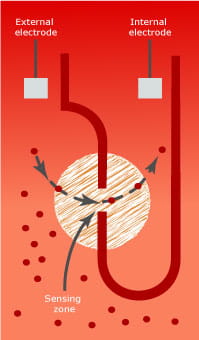
FIG. 3. The Coulter principle
Limitations:
- When a high reticulocyte or WBC count is nowadays, hematocrit determinations using hematology analyzers can event in the calculation of falsely elevated values, because the higher prison cell volumes of these cells volition interfere with the cerise blood jail cell count and the calculation of the hematocrit [vii]. See also Tabular array I.
three. Conductivity on blood gas analyzers
In POCT, blood gas analyzers measuring multiple STAT parameters are often used. Some claret gas analyzers determine hematocrit past a electrical conductivity measurement which is corrected for the concentrations of conducting ions in the sample.
Materials:
- Syringes or capillaries containing heparinized arterial or venous claret
Method:
- The electrical conductivity is the power of a solution to transmit (deport) electricity. The electrical electric current volition increase in proportion to the number of ions (or charged particles) found in a solution, their electric accuse and mobility, i.e. how hands the ions can move in the solution. The mobility of an ion in a solution will besides depend on how many cells (and size and shape) are suspended in the solution.
- Both erythrocytes and plasma take feature electrophysical properties. The membrane of the erythrocytes is electrically insulating, mainly due to its content of lipids, and so that information technology appears essentially non-conducting.
- Plasma is fairly conductive due to its content of electrolytes and charged proteins; the major contributor to plasma conductivity is Na+, the concentration in man blood plasma existence approx. 140 mmol/L.
- Due to this, at that place is an inverse relationship between the electrical conductance and the hematocrit in blood when the concentration of the charged particles is taken into account.
Three factors likewise the number of RBCs are critical for the conclusion of the hematocrit value when using a method based on measurement of electric electrical conductivity:
- Electrolytes
- Temperature
- Proteins
Most claret gas analyzers allow for these variables as follows:
- Concentration of electrolytes: This is determined past one or more ion measurements. Equally sodium is the main electrolyte in plasma, this is the about important ion to measure and use in the calculation of hematocrit.
- A change in the temperature has a meaning impact on the electrical conductivity because claret has a high temperature coefficient. The measuring chamber in blood gas analyzers is thermostatted and the blood sample preheated prior to measurement; thus there is no contribution from irresolute temperature.
- The protein concentration in plasma is causeless abiding in healthy people, so a constant compensation for this is incorporated in the adding of hematocrit on claret gas analyzers.
Limitations:
- In patients with abnormal plasma osmolality, e.thou. patients being treated with plasma expanders, claret diluents or massive infusion therapy, the protein concentration is no longer constant and the hematocrit conclusion gives falsely low values [8, 9]. Some blood gas analyzers offer correction for this bias [10,xi,12]. Come across likewise Table I.
4. Adding of hematocrit from hemoglobin
As there is a linear relationship between hemoglobin (ctHb) and hematocrit as described earlier, information technology is possible to summate the hematocrit on analyzers that measure hemoglobin. When making this conversion, two factors should be taken into consideration:
- The analytical quality of the ctHb measurement
- The precision of the equation that converts the two parameters
The measurement of ctHb from most expert-quality analyzers is unremarkably reliable; however, the equations used to calculate the hematocrit vary from analyzer to analyzer. Some analyzers use an empirically establish equation [2,xiii] whereas others utilize an approximate conversion factor of 3 [14,15].
Example:
| Hb concentration | Conversion equation | Hct % | Reference |
| xv* | Hct (%) = (0.0485 × ctHb (mmol/L) + 0.0083) × 100 | 45.98 | [2] |
| 15 | Hct (%) = ii.eight × ctHb (g/dL) + 0.8 | 42.80 | [xiii] |
| 15 | Hct (%) = ctHb (g/dL) / 0.34 | 44.12 | [xvi] |
| 15 | Hct (%) = 2.941 × ctHb (g/dL) | 44.12 | [xiv,fifteen] |
TABLE I: Effect of different conversion factors on Hct %.
* Conversion gene: yard/dL × 0.62058 = mmol/Fifty
Limitations:
- It is generally assumed that the conversion from hemoglobin to hematocrit is straightforward since most methods measuring ctHb are considered to be adequately authentic; however, different analyzers utilise dissimilar conversion factors, which may compromise the reliability of the hematocrit outcome. Hematocrit and hemoglobin are ofttimes used interchangeably; all the same, different studies have shown that the two parameters are not comparable, but that they accept their separate applications [15,17,18,19].
Comparing measuring technologies
All measuring technologies for determining hematocrit take advantages and disadvantages. The following tabular array provides an like shooting fish in a barrel overview of the described methods. The advantages/disadvantages listed have all been constitute past reviewing the literature, i.east. no prioritizing according to importance has been done.
Table as word document.
| Method | Advantages |
| Microhematocrit |
|
| Complete blood jail cell count |
|
| Electrical conductivity |
|
| Calculation from ctHb |
|
TABLE IIa. Advantages of different methods of measuring hematocrit.
| Method | Disadvantages |
| Microhematocrit |
|
| Consummate blood cell count |
|
| Conductivity |
|
| Calculation from ctHb |
|
TABLE IIb. Disadvantages of different methods of measuring hematocrit.
Discussion
When hematocrit is used to assess anemia and the oxygen-conveying capacity, the advantages and disadvantages of each method must be advisedly considered. In add-on, the specific clinical and analytical needs for any patient population must be determined.
Hematocrit is traditionally a routine hematology laboratory parameter; nevertheless, measuring hematocrit equally a STAT parameter in a POC setting is in many ways preferable, every bit this volition save fourth dimension in critical situations and avoid specimen send problems. Some claret gas analyzers offer this possibility, either by measuring hematocrit by electrical conductivity or past calculating hematocrit from ctHb.
Hemoglobin is as well used to appraise anemia both in the laboratory and as a STAT parameter on POCT analyzers. Dissimilar hemoglobin measuring methods and their applications volition be discussed in some other commodity.
Decision
Both POCT and traditional laboratory methods of measuring or computing hematocrit have limitations. Methods used outside of the laboratory must be intuitive for users without time-consuming sample handling.
Methods suitable for an adult environment may non exist suitable for a neonatal environment due to sample book limitations. Some methods are not suitable in sure applications due to e.chiliad. problems associated with book expansion fluids or abnormally sized or shaped red blood cells.
These limitations can have important clinical implications and must be carefully considered every bit described in this article.
macdonaldmurst1966.blogspot.com
Source: https://acutecaretesting.org/en/articles/hematocrit--a-review-of-different-analytical-methods
0 Response to "How to Read a Microhematocrit Tube Reader"
Postar um comentário